Efficient sensors for chemical warfare agents via experiment and #compchem

Chemical warfare agents put at stake human life and global safety. These compounds are extremely toxic, and their efficient detection is crucial.

By combining experiments and theory, we realized a new sensor, based on manganese oxide and gold nanoparticles, which has ultra-low detection limits and impressive selectivity towards an important simulant of the vesicant nitrogen mustard gas.

The manganese oxide nanomaterials were synthesized by chemical vapor-deposition (CVD), starting from a Mn(II) molecular complex. This compound can be easily vaporized, and gives manganese-oxide materials of high purity. Then, the manganese oxide surface was partially covered by gold nanoparticles.

Scheme of the synthesis of the Au-decorated manganese oxide nanomaterials

The material was then tested in the detection of a nitrogen mustard gas simulant (named dipropylene glycol monomethyl ether, DPGME). The results were exciting: the sensor showed high efficiency and selectivity, with a detection limit of 0.6 ppb.

All this work was done by our awesome experimental colleagues. However, I want to show you that also theoretical modeling (#compchem) has done its part here, by answering the question: how does this system work?

At molecular level, the sensing action depends on the contact of the analyte molecules with the active part of the sensor – the gold/manganese oxide layer. By using a density functional approach, we have seen that the molecule strongly binds to both gold and metal oxide, as shown in the picture below.

The mustard gas simulant DPGME (cyan,white,and red spheres) binds to both the Mn3O4, surface (ball-and-sticks) and gold (orange spheres). The top part of the figure shows the response of the chemoresistive sensor to the adsorbed DPGME molecules.

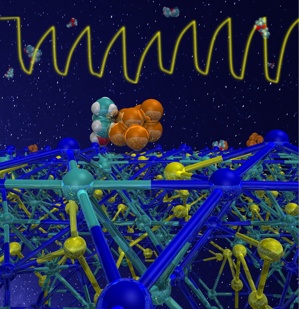
To check if this “molecular recognition” ability of the sensor was specific only to the target molecule, we modeled also the contact of ethanol with the sensor.

We found that while the mustard gas simulant is in intimate contact with both gold and Mn3O4, ethanol interacts only with the oxide surface, but not with gold. This can explain the higher response and selectivity of DPGME with respect to ethanol.

In short, the new materials have a low fabrication cost and remarkable sensing capabilities. The reason of their impressive performances in the detection of the mustard gas simulant is that the target molecule is anchored to both manganese oxide and gold.

I was really proud to present this work at the #RSCPoster2020 event – and I thank the organizers for this fantastic (and fun) opportunity.

By the way, if you still don’t know what a #RSCPoster conference is, follow #RSCPoster on Twitter, have a look to the many many great posters presented this year, and consider taking part to the 2021 edition!

Here’s our little contribution to this wonderful global event

Our poster presented at the 2020 #RSCPosterTwitter Conference

If you’d like more information this work – including the technical details of both modeling and experiments – these can be found in our published paper (see here for a free green open access version).

Water in hydrophobic and hydrophilic channels

Water. Always difficult to write something original about it, but let’s spend again a few words in celebration of this molecule. Water is present, or can be inserted in many porous hosts, like zeolites or MOFs. Not all of them love water. This time the question was: what does water do inside channels of similar size but different hydrophilicity?

We modelled the behaviour of water in two porous materials. The first one is zeolite L, which is hydrophilic. The second one is a metal organic framework, or MOF, which has pores of similar size, but less affinity to water. Our starting point was the X-ray structure of the two materials, shown below.

seminar_lausanne_fin23-e1563906626178.jpg
X-ray structure of  ZL-MOF and zeolite L, viewed perpendicular to the channel axis. In both materials, the water positions (cyan spheres) are partially occupied

The water distribution inside the pores looks very nice and symmetric. Unfortunately, the water positions are only partially occupied. So, by using the experimental water content as input, we optimized the structure of these materials, and here’s what we got.

seminar_lausanne_fin_zlmof3
Optimized structures of ZL-MOF (left) and zeolite L (right), viewed along the channel axis. The hydrophobic methyl groups of the MOF (in gray) protrude inside the channel and force the water molecules to arrange in well-separated rings. In the hydrophilic channels of zeolite L, a continous water structure is formed.

We found that water stabilizes both materials, and that the shape of the water clusters inside the channels depends on the affinity of the hosts to water.
While the hydrophobic host contains water rings, kept together by water-water hydrogen bonds, the hydrophilic host contains a continuous water tube, stabilized by interactions with the zeolite and also by hydrogen bonds.

seminar_lausanne_fin_zlmof2
Optimized structures of ZL-MOF (left) and zeolite L (right) (front view). While the water rings in the MOF are dominated by water-water hydrogen bonds, the water molecules in zeolite L can interact very strongly also with the potassium cations (purple spheres) and the framework.

In the zeolite channels, some water molecules are surrounded by five strong hydrogen bonds. This structure is similar to water pre-dissociation complexes found in liquid water, and it might probably explain the high proton activity found in zeolite L.

zlmofzl
Water rings in ZL-MOF (left), and water pre-dissociation complex (in red) in zeolite L (right)

Zeolite L is a promising material for solar cell applications, but the high proton activity inside the channels might damage some of the organic dyes that are incorporated as guests. Now we have identified a possible cause of the problem, and this might be a first step to improve the performances of these materials. Also, we hope that the atomistic insight on the water rings inside the MOF could help to exploit this material as host matrix for new compounds.

Personally, I much enjoyed doing this work: there’s always something to learn about confined water! The “driving force” for starting this work was an invitation, so many thanks to Michael Fischer and Robert Bell for organizing the Special Issue “Modelling Crystalline Microporous Materials” in the Zeitschrift für Kristallographie. If you like porous materials and #compchem, please have a look at this issue, it has many beautiful contributions.  Also, thanks to ChemRxiv for hosting our preprint,  and to the 2019 Twitter #RSCPoster conference, where this work was first presented as  poster.

The devil makes the pots but not the lids.

il diavolo fa le pentole ma non i coperchi

The title of this post is the literal translation of a proverb. The proverb means that Devil’s pot of wickedness sooner or later will boil – and, as there’s no lid, someone will see its content and reveal the truth. That’s the old innocent idea that, finally, justice will prevail over evil… well, I like it so much I use it as title. Rather than devils, this post is actually about pots and lids – of molecular size, of course.

As that’s not a Masterchef contest at the nanoscale, let’s get rid of the pot for the moment, and call it ‘container’. In the nanoworld there are many such containers, which can be filled with molecules. In this way, you can produce new materials with applications in various areas of technology: from solar energy to sustainability and human health.

Our containers are named zeolites – porous materials which are commonly used as adsorbents and catalysts in various commercial, industrial, and even medical applications as well as in our everyday life.  Also, if you fill zeolites with dye molecules, you’ll get materials able to capture and transfer solar energy very efficiently. You would do it much easier if you first know how their pores look like.

In particular, how do their entrances appear to an incoming molecule? This question is our “step one”,  because this information is really hard to get from experiments.

step01

Fortunately, modeling comes to the rescue…. and that’s one of the reasons why I love so much doing #compchem (computational chemistry)!!

step02

Step 2 revealed that the channel openings expose hydroxyl groups, and look somewhat like this:

zeoliteLChannel
Entrance of zeolite L channel, showing the terminal -OH groups and the channel accessibility.

Those terminal hydroxils can be condensed with other molecules, carrying specific groups, hence new properties and functionalities. Among them, the possibility of “closing” the pores. Why is it so important?

Zeolites are resistant to heat and pressure, and act as a protective shield around the dye. But every “pot” needs a “lid”:  plugging the zeolite pore entrances, so that the dyes, once included, cannot escape into the environment, would further enhance their stability.  This has already been done experimentally,  by attaching at the channel entrances peculiar molecules nicknamed “stopcocks”. They consist of two “parts”:

  • the “tail”, which can penetrate zeolite pores;
  • the “head”, which is too big to enter the pore and remains outside, thus blocking (at least partially) the channel opening.

Two typical stopcocks, one with a small tail, and the other with a long, bulkier tail, are shown below.

stopcoc

Such “molecular stoppers” do indeed a great job in preventing molecules to escape from zeolites.  However, there were no clear ideas about how these stoppers were attached to the pore entrance, and how much space they occupied.  This knowledge would help finding better “lids” for our zeolite “pots”. How do we get it? Of course by modeling, as sketched in step 3 and 4.

step03

Here’s what we learned:

  • stopper molecules prefer to bind aluminum sites at the channel entrance;
  • the tail group always penetrates inside the pore, while the head stays outside;
  • the extent of blocking depends on the stopcock.
    In particular:

     – small-tailed stopcocks are like partially opened “lids” : no full closure                – bulky-tailed stopcoks behave like “corks”: full closure

So the zeolite pore may be fully sealed by one bulky stopper, like a molecular cork on a Prosecco nano-bottle. On the contrary,  one “lid” (small stopper) leaves our “pot” partially opened. Fortunately, there’s enough room to attach a second small stopper to the opening, that can now fully be closed.

And this brings us to step 5…

step05

… which could well be the end of this story, first told some time ago. Thank you for reading it!

Anyway, there’s an epilogue, which is perhaps the nicest part (“dulcis in fundo“).  Using such information, obtained from modeling, experimental colleagues recently trapped indigo (that’s, your denim’s blue) in zeolite L, and blocked the channel entrances with two small stopcocks. In this way, they made a new pigment, exceptionally resistant, with an amazingly beautiful blue color.  For me #compchemist, that blue was simply….. the color of happiness.

zeolite_channel_openings


For more information…