The replacement of fossil fuel with sustainable alternatives free from environmental footprint is one of the most important challenges to combat climate change and meet the ever increasing energy demand of our planet. The sustainable production of hydrogen fuel through biomass-derived ethanol in Direct Ethanol Fuel Cells is a promising route, but the high costs and short lifecycle of platinum – which is still the preferred catalyst – are a serious problem. So, the quest to valid yet convenient substitutes to platinum is an open and challenging task.
We (actually, my experimental colleagues) prepared an electrocatalysts for Ethanol Oxidation Reaction based on a low-cost and abundant metal oxide, namely manganese oxide. The fabrication strategy involves the growth of manganese oxide nanostructures on nickel foam scaffolds via plasma-assisted chemical vapor deposition and the functionalization with gold nanoparticles in low amount – as sketched in the picture below. That’s the magic of molecule-to-nanomaterials conversion!
The synthesized nanostructures have large surface area and show great performances as electrocatalysts in the ethanol oxidation reaction, comparing favourably with the best oxide-based catalysts known to date. We found that a very tiny amount of gold nanoparticles is sufficient to boost the catalytic activity of manganese oxide.
Our findings not only afford a convenient route for sustainable electrocatalysts, but also explain why our catalyst is so efficient. Theoretical modeling (#compchem) showed that gold nanoparticles activate the oxide surface toward the ethanol oxidation reaction. In other words, ethanol undergoes both partial oxidation and deprotonation immediately upon adsorption on the catalyst. Hence, our catalyst optimally prepares ethanol to the electrochemical oxidation process.
This knowledge, combined with the proposed fabrication route, may guide the development of electrocatalysts based on earth-abundant metal-oxides for ethanol valorization in Direct Ethanol Fuel Cells and for (photo)electrochemical water splitting.
Personally, I enjoyed very much this work, because metal-metal oxides interfaces are particularly challenging to deal with by #compchem. Also, I like very much to interact with my experimental colleagues and friends: they always have interesting problems, and collaborating together to find a solution is often the best part of the work. Very happy that computational modeling may help to understand the complex behaviour of these intriguing materials!
We presented this work at the fabulous #RSCPoster conference 2021. Here’s a pdf copy of our poster.
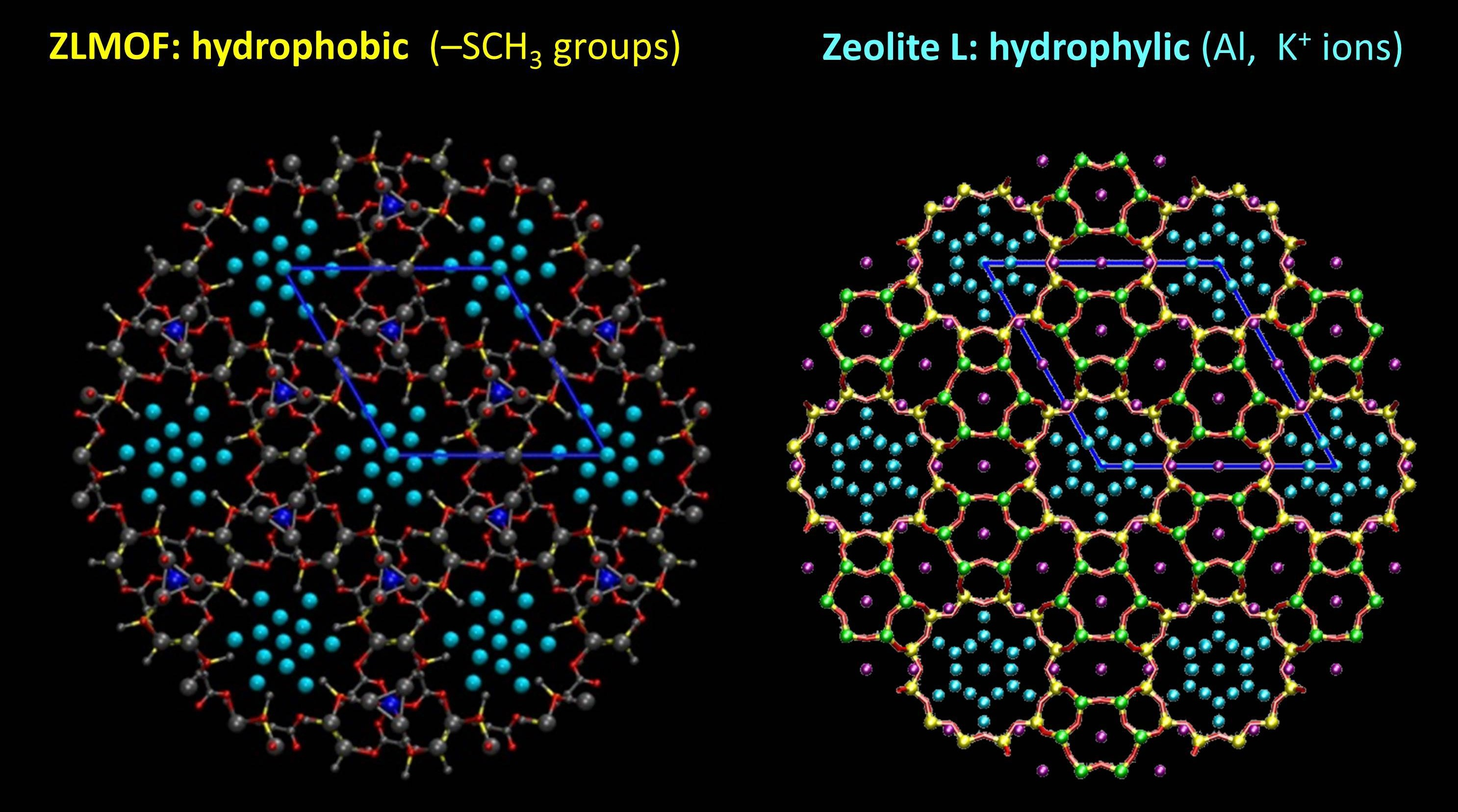
Water. Always difficult to write something original about it, but let’s spend again a few words in celebration of this molecule. Water is present, or can be inserted in many porous hosts, like zeolites or MOFs. Not all of them love water. This time the question was: what does water do inside channels of similar size but different hydrophilicity?
We modelled the behaviour of water in two porous materials. The first one is zeolite L, which is hydrophilic. The second one is a metal organic framework, or MOF, which has pores of similar size, but less affinity to water. Our starting point was the X-ray structure of the two materials, shown below.
X-ray structure of ZL-MOF and zeolite L, viewed perpendicular to the channel axis. In both materials, the water positions (cyan spheres) are partially occupied
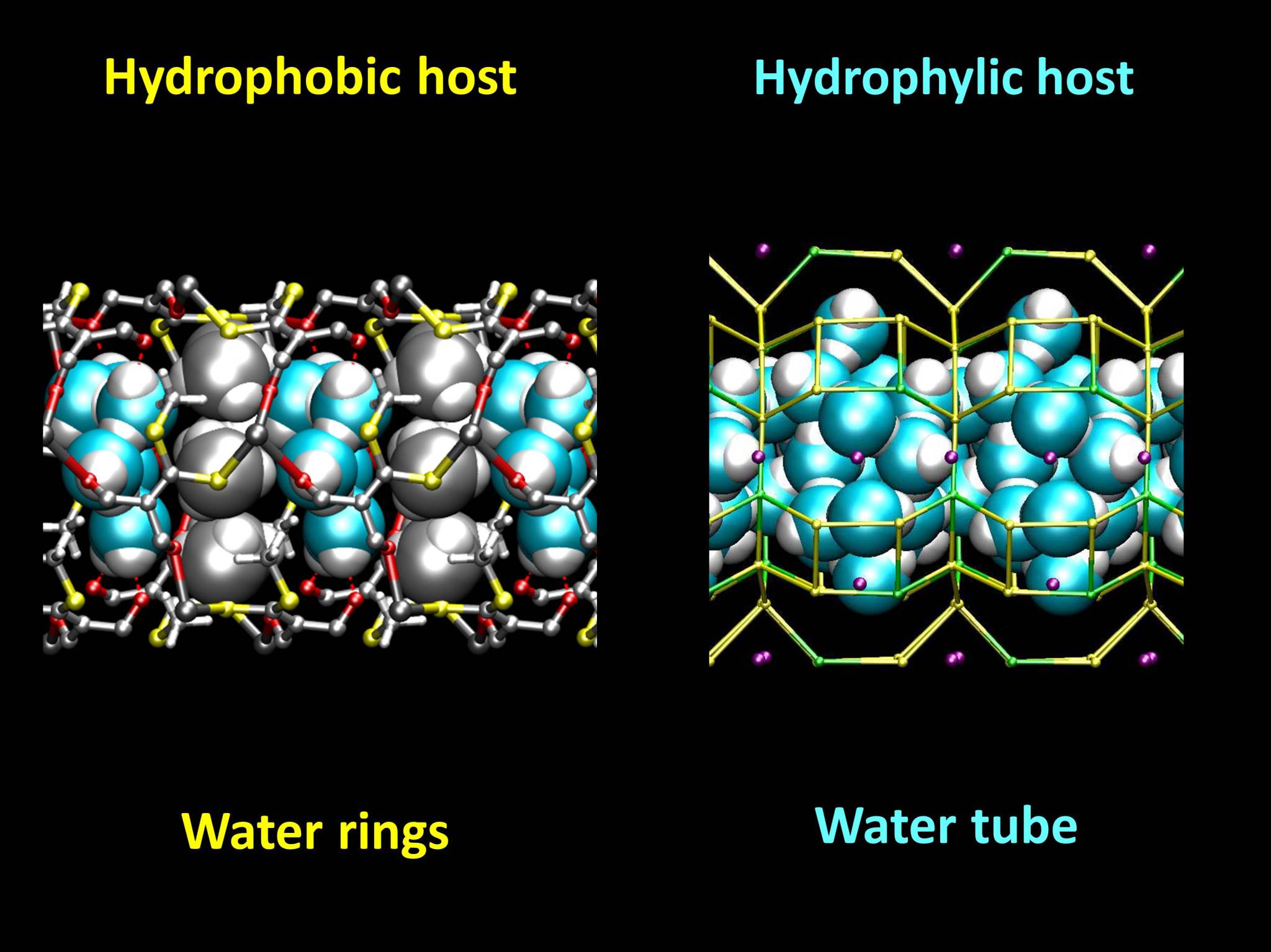
The water distribution inside the pores looks very nice and symmetric. Unfortunately, the water positions are only partially occupied. So, by using the experimental water content as input, we optimized the structure of these materials, and here’s what we got.
Optimized structures of ZL-MOF (left) and zeolite L (right), viewed along the channel axis. The hydrophobic methyl groups of the MOF (in gray) protrude inside the channel and force the water molecules to arrange in well-separated rings. In the hydrophilic channels of zeolite L, a continous water structure is formed.
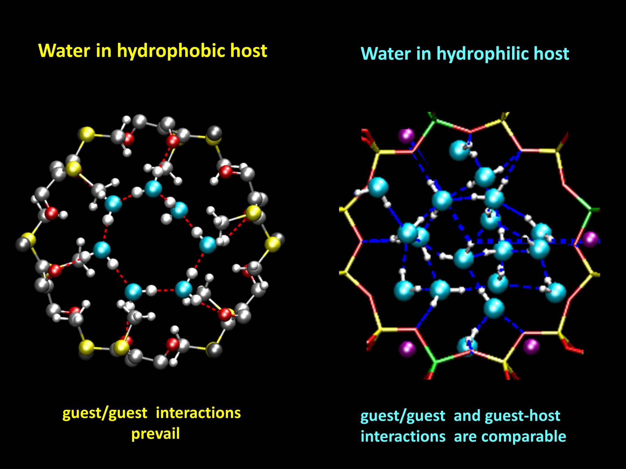
We found that water stabilizes both materials, and that the shape of the water clusters inside the channels depends on the affinity of the hosts to water.
While the hydrophobic host contains water rings, kept together by water-water hydrogen bonds, the hydrophilic host contains a continuous water tube, stabilized by interactions with the zeolite and also by hydrogen bonds.
Optimized structures of ZL-MOF (left) and zeolite L (right) (front view). While the water rings in the MOF are dominated by water-water hydrogen bonds, the water molecules in zeolite L can interact very strongly also with the potassium cations (purple spheres) and the framework.
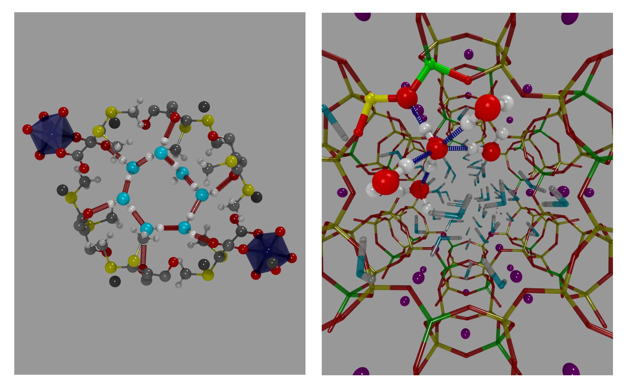
In the zeolite channels, some water molecules are surrounded by five strong hydrogen bonds. This structure is similar to water pre-dissociation complexes found in liquid water, and it might probably explain the high proton activity found in zeolite L.
Water rings in ZL-MOF (left), and water pre-dissociation complex (in red) in zeolite L (right)
Zeolite L is a promising material for solar cell applications, but the high proton activity inside the channels might damage some of the organic dyes that are incorporated as guests. Now we have identified a possible cause of the problem, and this might be a first step to improve the performances of these materials. Also, we hope that the atomistic insight on the water rings inside the MOF could help to exploit this material as host matrix for new compounds.
Personally, I much enjoyed doing this work: there’s always something to learn about confined water! The “driving force” for starting this work was an invitation, so many thanks to Michael Fischer and Robert Bell for organizing the Special Issue“Modelling Crystalline Microporous Materials” in the Zeitschrift für Kristallographie. If you like porous materials and #compchem, please have a look at this issue, it has many beautiful contributions. Also, thanks to ChemRxiv for hosting our preprint, and to the 2019 Twitter #RSCPoster conference, where this work was first presented as poster.
Overwhelmed with the increasing flow of new scientific discoveries and related literature? You’re not alone. We live in the information overload era: too much to read, too little time, and life is short. Probably we’d need more readable, shorter papers too. Why writing a long one? Perhaps, it might connect disciplines which speak different languages but have much in common. Like material science and mineral science.
Let’s start from the first one.
You can make materials for solar cells, optical devices or medical sensors by trapping molecules or nanoparticles inside a “host”. Once there, molecules are no longer free to move, like in a gas or a liquid. This process, called “confinement”, brings to life new properties, which were not present in the individual molecules and are very useful in technology. Energy transfer or information storage, for instance, are made possible by the organization of the confined molecules.
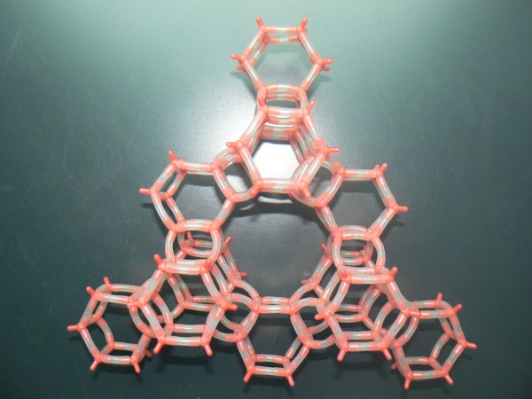
The regular cavities of zeolites do a great job in organizing guest molecules
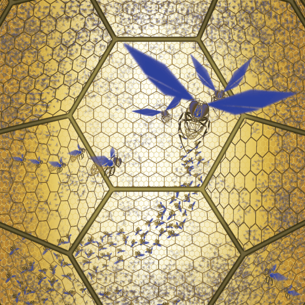
Tiny smart objects such as molecular machines, motors and diodes, make good use of self-organization processes, which create order from apparent disorder by exploiting interactions between molecules. This task gets easier when molecules are confined in regular pores. Think of a buzzing swarm of bees, first frantically hovering in the air, and then accommodated in a honeycomb.
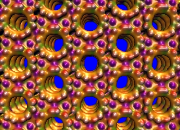
Similar to honeycombs, regular patterns of pores like those in zeolites can orderly accomodate small molecules or clusters. But if you want to entrap, say, enzymes, peptides, or large nanoparticles, you must use materials with larger pores. Some porous silicas have large honeycomb channels, while the cavities of metal organic frameworks display an amazing variety of size and shape. With those nice architectures awaiting to be filled, ordering molecules might appear like an easy task.
As you imagine, things are more complex. Perfect order cannot be achieved. All cavities would need to be uniformly occupied by the guests. This is going to be very unlikely, because molecules move a lot even when they’re confined… like bees in a hive.
Molecules in nanocavities are sort of like bees in a honeycomb: they form an organized colony (Artwork: Andrea Stangoni)
About bees, I had direct experience… as a child, I used to observe my dad opening up his hives to inspect them. This gave me the chance to “study” the behaviour of these awesome creatures inside their honeycomb.
Bees do not occupy all hexagonal holes in the frame, and move continuously around, without any apparent pattern. Hence they’re not perfectly ordered. In spite of this, the colony is amazingly organized, and performs an impressive number of complex tasks…. not just honey production!
Similarly, guest molecules confined in porous cages are not rigorously ordered. Yet they are organized, and the resulting host-guest materials can perform useful functions, which were absent in the free molecules. They can, for example, absorb and transfer photons like the antenna systems of plants and bacteria.
Now, the question is: can we improve the organization of the molecules and the performances of the materials? Well, first we should know how the molecules occupy the cavities, their orientation, spacing and so on. Are the guests aligned? Are they attached to the pore walls? What happens if water enters the pores? To find those answers, you should use several different techniques: each experiment will give you some pieces to compose the puzzle. And yes, computational chemistry helps a lot to figure our what happens inside the pores. Yet this remains a very difficult problem.
This is where mineral science might help.
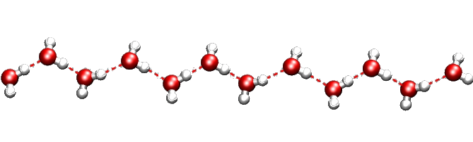
Regular patterns of cages are very common in the mineral world. Not long ago, for example, geologists found in Antartica a mineral with the same structure of zeolite Z-SM5, a well-known and widely used artificial industrial catalyst. That was indeed a big surprise! Natural zeolites are indeed amazing: their pores contain impressively stable structures formed by small molecules and cations. Just look at this water wire:
Water wire found in the channels of a natural zeolite
Contrary to what you’d expect, this chain is incredibly resistant to heat and pressure. First found in a rare mineral, it was named “one-dimensional ice”. But actually, our water wire “melts” at about 340 C inside the mineral framework! This is a great example of organized structure made by Nature. You can find many others: the most famous ones are perhaps gas hydrates. Several silica minerals have hydrate structures, which are also very common in man-made porous materials. Indeed, we should pay more attention to the close links between natural and artificial host-guest materials.
Natural porous minerals, the intriguing organization of their guests, and their response to mechanical stress can be an awesome source of inspiration in the quest of more robust and efficient materials. High pressure experiments with zeolites (and also some MOF’s) have already brought us new organized materials, along with many curious facts. But there’s so much yet to be discovered.
Perhaps, the problem with us (me included) and with our scientific era is that we don’t take enough time to relate with other disciplines. I’ve been so lucky to work with many awesome colleagues from the mineral, chemical and material science communities over the years, and it’s thanks to them that I wrote this review. One thing I learnt is that we should always try building bridges and strenghtening links between different fields because there’s nothing to lose, all to gain from a deeper exchange of ideas.
The title of this post is the literal translation of a proverb. The proverb means that Devil’s pot of wickedness sooner or later will boil – and, as there’s no lid, someone will see its content and reveal the truth. That’s the old innocent idea that, finally, justice will prevail over evil… well, I like it so much I use it as title. Rather than devils, this post is actually about pots and lids – of molecular size, of course.
As that’s not a Masterchef contest at the nanoscale, let’s get rid of the pot for the moment, and call it ‘container’. In the nanoworld there are many such containers, which can be filled with molecules. In this way, you can produce new materials with applications in various areas of technology: from solar energy to sustainability and human health.
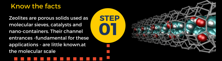
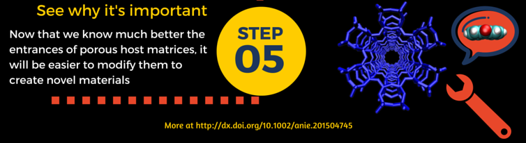
Our containers are named zeolites – porous materials which are commonly used as adsorbents and catalysts in various commercial, industrial, and even medical applications as well as in our everyday life. Also, if you fill zeolites with dye molecules, you’ll get materialsable to capture and transfer solar energy very efficiently. You would do it much easier if you first know how their pores look like.
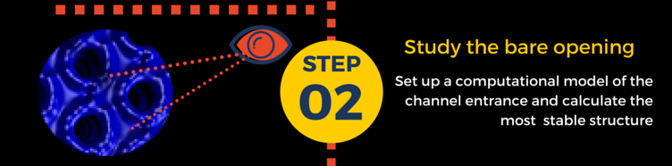
In particular, how do their entrances appear to an incoming molecule? This question is our “step one”, because this information is really hard to get from experiments.
Fortunately, modeling comes to the rescue…. and that’s one of the reasons why I love so much doing #compchem (computational chemistry)!!
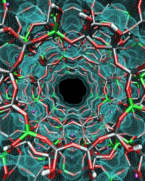
Step 2 revealed that the channel openings expose hydroxyl groups, and look somewhat like this:
Entrance of zeolite L channel, showing the terminal -OH groups and the channel accessibility.
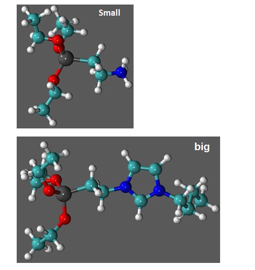
Those terminal hydroxils can be condensed with other molecules, carrying specific groups, hence new properties and functionalities. Among them, the possibility of “closing” the pores. Why is it so important?
Zeolites are resistant to heat and pressure, and act as a protective shield around the dye. But every “pot” needs a “lid”: plugging the zeolite pore entrances, so that the dyes, once included, cannot escape into the environment, would further enhance their stability. This has already been done experimentally, by attaching at the channel entrances peculiar molecules nicknamed “stopcocks”. They consist of two “parts”:
the “tail”, which can penetrate zeolite pores;
the “head”, which is too big to enter the pore and remains outside, thus blocking (at least partially) the channel opening.
Two typical stopcocks, one with a small tail, and the other with a long, bulkier tail, are shown below.
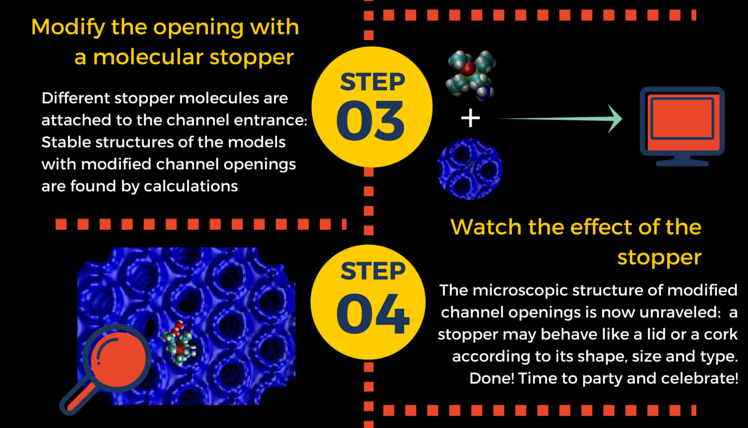
Such “molecular stoppers” do indeed a great job in preventing molecules to escape from zeolites. However, there were no clear ideas about how these stoppers were attached to the pore entrance, and how much space they occupied. This knowledge would help finding better “lids” for our zeolite “pots”. How do we get it? Of course by modeling, as sketched in step 3 and 4.
Here’s what we learned:
stopper molecules prefer to bind aluminum sites at the channel entrance;
the tail group always penetrates inside the pore, while the head stays outside;
the extent of blocking depends on the stopcock.
In particular:
– small-tailed stopcocks are like partially opened “lids” : no full closure – bulky-tailed stopcoks behave like “corks”: full closure
So the zeolite pore may be fully sealed by one bulky stopper, like a molecular cork on a Prosecco nano-bottle. On the contrary, one “lid” (small stopper) leaves our “pot” partially opened. Fortunately, there’s enough room to attach a second small stopper to the opening, that can now fully be closed.
And this brings us to step 5…
… which could well be the end of this story, first told some time ago. Thank you for reading it!
Anyway, there’s an epilogue, which is perhaps the nicest part (“dulcis in fundo“). Using such information, obtained from modeling, experimental colleagues recently trapped indigo (that’s, your denim’s blue) in zeolite L, and blocked the channel entrances with two small stopcocks. In this way, they made a new pigment, exceptionally resistant, with an amazingly beautiful blue color. For me #compchemist, that blue was simply….. the color of happiness.
Requiring subscription: i) Indigo in the nanochannels of zeolite L (Woodtli et al, Dyes and Pigments, 2018, 149, 456); ii) Invention of the stopcock (Maas & Calzaferri, Angew Chem 2002, 41, 2284); iii) Official version of our article: Structure of Nanochannel Entrances in Stopcock‐Functionalized Zeolite LComposites,Angewande2015, 54, 11112
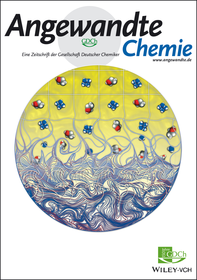
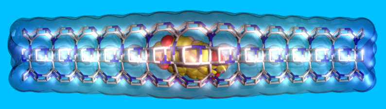
When we fill porous materials with dye molecules of the right size, we obtain useful compounds for solar energy technology. These compounds can transfer solar energy efficiently because pores and channels fit to the dyes “like a glove”. In this way, molecules are forced to stay in line, and energy can easily pass from a molecule to the next one in the line. If we knew in detail the structure of the dye arrays, we’d have better chances to improve these compounds.
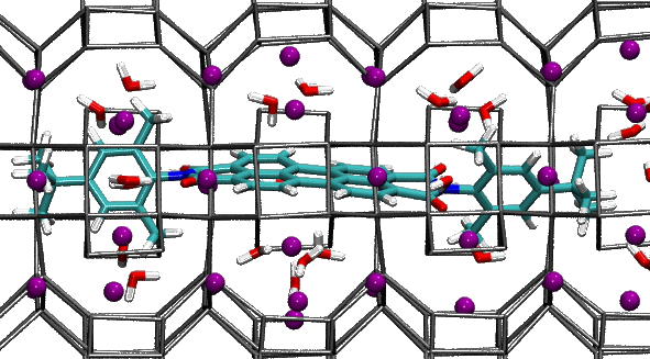
Unfortunately, the precise positioning of the molecules inside the pores is very hard to determine. Recently, we solved this problem for a class of particularly efficient dyes filling the channels of zeolite L. Key to success was diversity within the team, which favored the combination of multiple techniques involving both experiments and calculations.
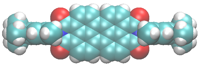
The useful properties of these materials arise from the arrangement of dye molecules inside the porous host, which depends on the interactions among molecules and with the porous host. After this work, now it seems we understand a little better these complex materials. Indeed, our dyes are linear, symmetric and fit to the zeolite channels. Yet they adopt a slightly asymmetric positioning to maximize the interactions with the zeolite cations, which stabilize the compound.
Perylene-bisimide dye (cyan) in zeolite L (gray). The purple spheres represent the zeolite potassium cations
This work also suggests some possible ideas to improve these compounds by modifying either the porous container (the “host”) or the dye molecule (the “guest”). In my view, this is also a good example of how computational modeling may help to rationalize experimental results in apparent contrast with each other, yielding a consistent picture of a useful and intriguing material.
Gigli et al. (2018) “Structure and Host–Guest Interactions of Perylene–Diimide Dyes in Zeolite L Nanochannels”J. Phys. Chem. C 122, 6, 3401-3418
On March 20th, i took part to the RSC Twitter Poster Conference 2017, an online event organized by The Royal Society of Chemistry to favour new contacts and exchanges among researchers in chemical sciences. The event was a big success.
To those of you that might wonder what a twitter poster session is, here’s an excerpt from The Analytical Scientist:
How do Twitter poster sessions work?
Participants tweet an image of their poster with the title and hashtags #RSCPoster and the area (e.g. #RSCAnal) at any point throughout a 24-hour period. This means that people anywhere in the world can join in.
It’s a fully global event open to every chemist on twitter. No conference fees: by following the hashtag #RSCPoster, anyone could attend and submit their poster.
What a nice surprise when nice images of posters started appearing in the feed on that Monday morning. Awesome idea – i thought, tweeting my contribution a few seconds later.
To be honest, the poster was not prepared for the occasion – I simply recycled a poster presented at a traditional conference, and I shared it just to see what would happen. It was great. Not only people were tweeting their images, they were also commenting on the posters, just like in a standard conference but within 140 characters. The participants were discussing technical aspects of the results or methodology, asking more general questions on the featured research, and all of this worked wonderfully. It was exciting: every few seconds, a new contribution was added to the feed, containing interesting and well presented science.
As in normal conferences, this wasn’t just a chance to present your own project, but also a fantastic opportunity to look at what the researchers out there were doing, and to learn a lot from it. New ideas were inspired by work in apparently unrelated research areas. Beside science, it was also a very useful experience in communicating research to a heterogeneous audience using few, carefully selected words. Yet another demonstration of how twitter can be useful to scientists!
I regret that i wasn’t able to look at all the posters during the session – they were definitely too many. Fortunately, even if the conference is over, the posters are still hanging on the virtual wall at the RSC Tumblir site, so that in the case you missed the event, you might still catch up with the interesting science. I’d strongly recommend to give a look at them: they’re awesome!
Some lucky participants got the coolest thing you could ever imagine: a cartoon abstact of their poster – like this one:
So I’m very grateful to @MCeeP (ErrantScience.com) for making my day with this, and for the incredible tour-de-force of drawing the cartoons! I much enjoyed to see all of them: not only they were funny, but also further engaged the participants, stimulating curiosity and new conversations. These brilliant poster abstracts really made the conference unique.
This is crazy but …what if cartoon abstracts were introduced in traditional conferences as well? To get a feeling, just check out the complete gallery of these cool cartoons of posters at ErrantScience.
Finally, many thanks to the RSC, the organizers, and the participants for such a great experience. A superb way to promote chemical research. I’m glad to have been some little part of it, and looking forward for the next year event.
Just for the records, here’s my humble contribution to #RSCposter. For those interested, what featured in the poster is briefly explained here (left side) and blogged here (right side).
Our work on ethanol and water in ferrierite, published here and blogged in my previous post, has been recently covered by MRS Bulletin in an excellent news article – “High pressure and small spaces create order from disorder” by science writer Tim Palucka. Some time ago, I had a very pleasant communication with Tim about the main ideas and results of the paper. That interview also helped me a lot to understand how science communication is done professionally. The piece by Tim really does a great job in explaining the scientific background, the main findings and the perspectives of our research – and, of course, all of us are so happy about it!
MRS Bulletin contains other interesting news articles, which are very useful to get a first impression about what’s going on in the many diverse areas of materials science – we’re very proud to be featured there! Big thanks, therefore, to MRS Bulletin and Dr. Palucka for the awesome coverage, and to Prof. Gion Calzaferri for commenting on our work as an external expert. A pdf version of the news article is freely available at MRS Bulletin (Volume 42, Issue 3, pp. 176-177, DOI: https://doi.org/10.1557/mrs.2017.38 ), while the illustration showing the arrangement of water and ethanol in the zeolite is just here below:
What happens to a liquid mixture when it is driven by pressure into an initially empty container? What if the container has an ordered pattern of molecular-sized pores? To answer this question, we prepared a sort of good vodka drink – three parts water, and one ethanol – and we injected it into the pores of an hydrophobic container – the zeolite ferrierite. As hydrophobic materials don’t like water and don’t care about drinks, we had to be very drastic: we used a diamond anvil cell. In this apparatus, the sample – the empty container and the mixture, in our case – is compressed between the tips of two opposing diamonds and experiences huge pressures – about 10.000 times the normal atmospheric pressure. At these conditions, matter is subjected to unimaginable forces, comparable to internal atomic forces: this means that strange, unexpected phenomena could show up. Now, let’s combine the power of high-pressures with the ordering effect of the pore matrix and see what happens to our mixture.
Just to start with, the water-ethanol mixture – the pressure-transmitting medium – enters the pores of the matrix. But how do the molecules occupy the pores? You don’t need to be a chemist to know how it is difficult to separate alcohol from water. This is a critical issue also for sustainable processes – such as the production of biofuels.
Thanks to high pressure and to the porous matrix – and with the help of computational modeling – here we obtained the separation of ethanol from water, and the formation of a beautiful pattern of clusters. The clusters – rows of ethanol dimers, and square water tetramers – occupy different regions of the host matrix and alternate like tiles forming a nice molecular mosaic – a “two-dimensional architecture” – inside the porous host. What’s really exciting about it is that the ordered pattern, created by high pressure, also remained stable by bringing the material back to atmospheric pressure. This means that using high pressures and porous hosts, we can create new materials, which are stable at normal conditions, and could potentially be exploited in applications.
The metamorphosis of the initial water-ethanol solution into a beautiful two-dimensional pattern remains somewhat mysterious. More in general, how organization arises from chaos is still one big question in science. However, our molecular dynamics simulations show that water molecules, already inside the pores, can spontaneously self-organize in square tetramers:
The final result, is the formation of the stable two-dimensional architecture of water and ethanol clusters. As the movie shows, the molecules move, but the clusters do not break apart. – even upon returning to room pressure.
Perspectives
Disclosing the way in which molecules and nanoparticles assemble at high pressure conditions, under the guidance of a suitable matrix would be a great and intriguing challenge for future studies. Another one would be the actual production of technologically relevant materials through the combined use of pressures and suitable porous matrices. These goals could be achieved only through a close collaboration between experiment and theory – a synergy which has been at the very origin of the present work.
In a wider perspective, understanding the behavior of matter at high pressures is of central relevance in science, as explained in this excellent introductory feature article. Pressure effects are ubiquitous, in chemistry, physics, earth and planetary sciences, as well as in many industrial processes and technological applications. High-pressure conditions are also hypothesized to explain the origin of complex chemistry and life. The study of this exotic regime, so different from our everyday-life, may reveal plenty of phenomena which would be hard to imagine based on our experience.
Reference: Irreversible Conversion of a Water–Ethanol Solution into an Organized Two-Dimensional Network of Alternating Supramolecular Units in a Hydrophobic Zeolite under Pressure, by Rossella Arletti, Ettore Fois, Lara Gigli, Giovanna Vezzalini, Simona Quartieri, and myself. Angewandte Chemie 2017 – DOI: http://dx.doi.org/10.1002/anie.201610949 http://dx.doi.org/10.1002/ange.201610949
Special thanks to Andrea Stangoni (@andrea_stangoni), author of the cover artwork. His image summarizes the ideas of our work much more beautifully than my blog post!
How can a snake swallow a mouse bigger than its mouth?
Weird as it seems, questions like this emerge very often at the molecular scale. For example, we can fill porous materials with molecules larger than the diameter of the pores: in this way, we may obtain devices for energy and health applications. What makes this useful process possible? Flexibility is the key: both the porous host (the “snake”) and the molecule (the “mouse”) must deform for the process to occur. But here, contrary to the mouse-snake case, cooperation between the two partners is needed.
We captured the passage of a bulky molecule through the very narrow opening of one of these pores. We did this by computer simulations, because it is very hard to get such information experimentally. To get an idea of what we found, you don’t even need to read the paper – and i’m not kidding. Just look at the movie below!
What we’ve seen first, is that the pore is slightly larger at its entrance. This surely helps the molecule to go in.
Second: contrary to the mouse, which would escape the snake as fast as it could, the molecule is indeed “magically” drawn to the pore entrance – by electrostatic forces.
“So what?” – you may say.
Keep in mind that the molecule is still larger than the pore opening. No kind of “fatal attraction” could do the trick, in a world of rigid bodies.
We’ve found that the molecule can pass through the opening and slip inside the pore only because it’s flexible, and its motion is “in tune” with the vibrations of the porous matrix. All this factors cope to make the entrance process more favorable than the exit process – that’s why the molecule gets finally swallowed by the pore, and remains trapped inside the material.
For me, it was very nice to see how bulky molecules manage to pass through narrow openings and travel inside a porous material. But finding out the reason why they stay inside was, probably, even more exciting: because it explains how materials of this kind can form and remain stable. Which is exactly one of the things you may need, in the quest of easier and smarter ways to produce better materials.
As we have to give credit where credit is due, i must confess that i borrowed the mouse-and-snake idea used in this post. But you’ll never know from whom. Me neither: (s)he was an anonymous referee of the paper. I am very grateful to this person: i can hardly imagine a nicest way to sketch our work.
Update:
Many thanks, of course, also to ChemComm for the cover!
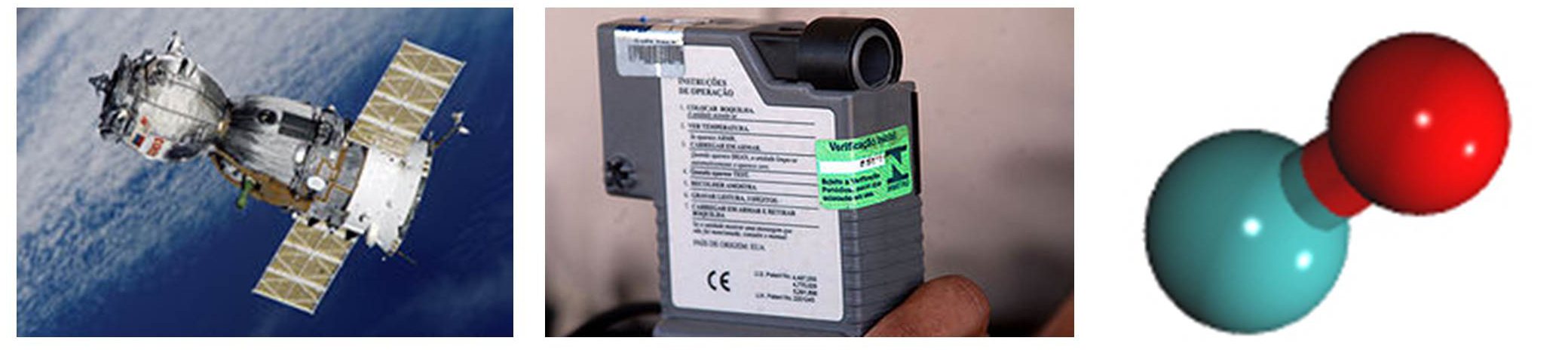
What do a spacecraft, a breathalyzer, and carbon monoxide have in common? Nothing at all – you’d think. And you’d be wrong! All three give you information on things that you cannot directly see, touch or measure. A spacecraft can capture some signal and send you beautiful images of a planet. With the help of a breath tester, a policeman may deduce the alcohol content in your blood. And using carbon monoxide, researchers may find highly reactive centers on materials surfaces. Let’s focus on the latter and see how it works!
Left image: the Soyuz spacecraft (source: Wikimedia commons). Center image: a breathalyzer (source: photograpy by Elza Fiúza/ABr, distributed under a CC-BY 3.0 license). Right image: carbon monoxide (blue=carbon; red=oxygen).
When a molecule comes in contact with surface atoms, its properties change. By measuring these changes, you get information on the surface sites interacting with the molecule. Molecular vibrations – that you can measure by infrared spectra – provide very useful information: the vibration of carbon monoxide is very sensitive to the type of surface sites. That’s why this molecule is used to identify active centers on catalytic materials, such as titanium dioxide.
How does carbon monoxide (CO) bind to surface atoms? If you’re a chemistry student, you (should) know very well how CO interacts with molecules and ions. You’ve learned that this molecule can work both as a donor and as an acceptor of electron density. Well, what’s nice, is that this happens also on surfaces, and you can see it experimentally.
Let’s see this step-by-step. Carbon monoxide is a peculiar molecule. When it acts as a donor, charge flows to its bonding partner, which could be, for example, a metal cation. This process strengthens the C−O bond and increases its vibration frequency. This means that, in the infrared spectrum of the sample, you’ll find the CO band at higher frequencies – “blue-shifted” – with respect to the free, unperturbed molecule. But carbon monoxide can also accept electron density from its bonding partner. If this occurs, the C-O bond becomes weaker: its stretching frequency decreases, and you’ll see a “red-shifted” CO band in your spectrum.
Carbon monoxide is colorless, odorless, and highly toxic – a true and unmerciful silent killer. It binds to iron(II) in hemoglobin, and this prevents the delivery of oxygen to the human tissues. This is a – very unfortunate – case where carbon monoxide acts at the same time as a donor and as acceptor. The bond is synergic: CO donates to the metal, the metal back-donates to CO, and these two mechanisms reinforce each other:– that’s why it kills. This synergy occurs in many molecular complexes of transition metals and ions – often with less dangerous consequences. It’s less known on metal oxide surfaces, but it may happen as well. Is this the case of TiO2?
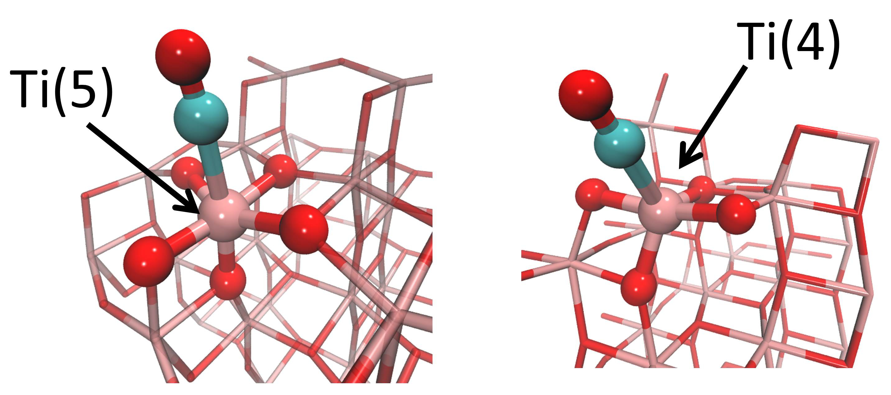
Not apparently, because carbon monoxide can only be a donor towards Ti cations – they are Lewis acids, and cannot give back electron density. The lower is their coordination number, the stronger is their acid power. For example, a Ti cation coordinated by 4 oxygens – Ti(4) – should be a stronger acid than one bound to 5 oxygens -Ti(5).
Researchers use carbon monoxide to explore the activity of surface cations and deduce their environment, in particular the number of oxygen neighbors. This information connects the reactivity of a catalytic center to its molecular structure, and may help them to improve the catalyst. Practically speaking, they send carbon monoxide on a TiO2 sample and measure the infrared spectrum. The rule is simple: the higher the frequency of the CO band, the more reactive are the Ti sites on the sample, and the lower their coordination number.
The image shows a 5-coordinated Ti center (left) and a 4-coordinated Ti site (right) on anatase-TiO2 surfaces
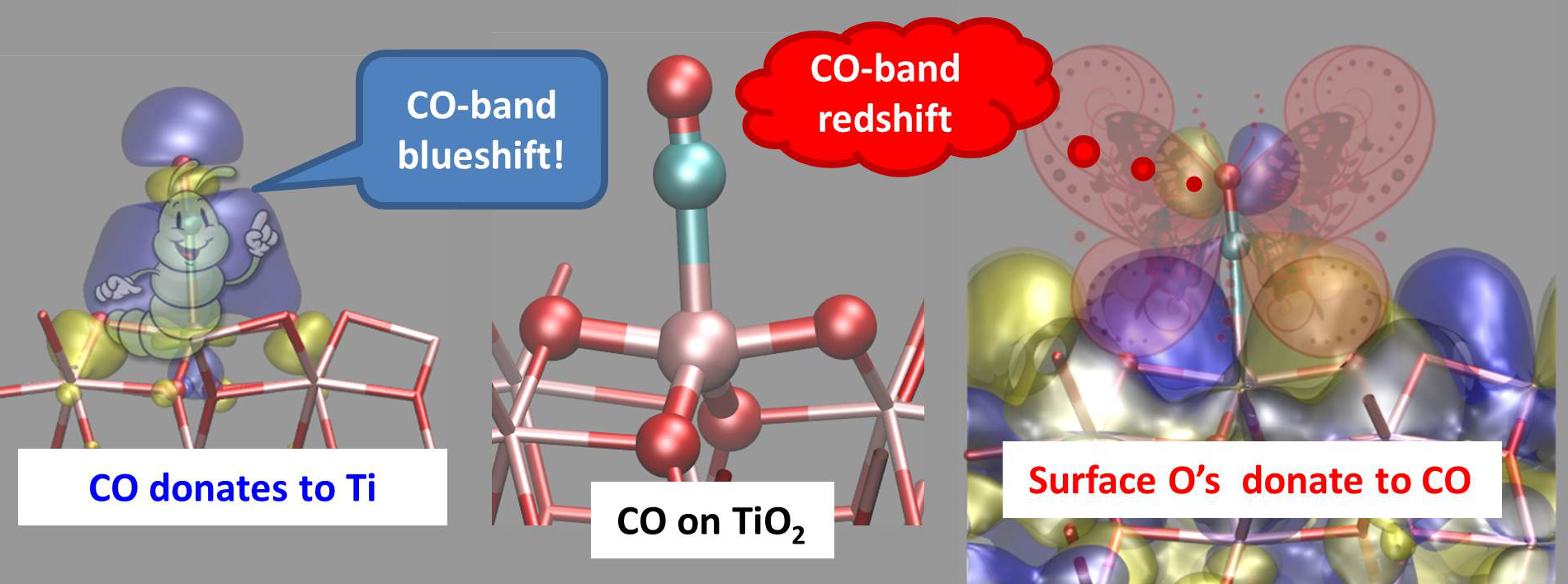
So if you had a TiO2 sample with Ti(5) sites, and a second one with Ti(4), what would you get from the experiment? “The second sample should show a more blue-shifted CO band, because Ti(4) is a stronger Lewis acid”. If you answered this, you’d be wrong… because we actually did the experiment, checked with calculations, and found the contrary. We found that CO on Ti(4) gives a less blue-shifted band – even if Ti(4) is a stronger Lewis acid. Just as if a breathalizer estimated a lower alcohol content in a drunker driver. This could happen only if a sort of magic potion neutralized the effects of alcohol (something similar exist in real life, but it’s a mineral and belongs to the large family of zeolites). Similarly, our carbon monoxide on Ti(4) should have received an antidote against the loss of electron density. The antidote could only be electron density: but where did it come from? Simply from the oxygen atoms bound to Ti(4): they are close enough to CO and ready to help.
In short, what happens is that CO donates electron density to Ti, but the surface oxygens donate electron density to CO. The first process strenghtens the C-O bond, but the latter has opposite effects. As a result, you find the CO signal at frequencies lower than expected. The two mechanisms are sketched in the figure below – my attempt to explain in a simple way the two-fold nature of the Ti-CO bond on titanium dioxide surfaces.
So, if you see high frequency bands in an infrared spectra of CO, please be warned: not necessarily they are due to very reactive sites on TiO2 surfaces. And also keep in mind that carbon monoxide gives you indirect information on your sample. Its signal can be influenced in complex ways by several factors – you might misinterpret your data, based on simple rules. From a practical viewpoint, i think that you should be aware of this, especially if you’re working on CO, or titanium dioxide materials. More speculatively, this story might help us to better understand how molecules interact with surface atoms. The complex, delicate balance of molecular-scale interactions is at the origin of technologically important phenomena – reactivity, catalysis, photocatalysis, just to mention some of them. Understanding these interactions more deeply could help us to improve their practical applications. Much effort is still needed, but it’s worth doing!
This research by our group has been published recently (Deiana et.al., ChemPhysChem2016, 17, 1956; 10.1002/cphc.201600284). It was also sketched in a short summary, and by an infographics in a previous post. Here i used other words to tell the same story, because i feel it’s important to make research results accessible to a larger community.


