Porous materials, minerals and bees

Overwhelmed with the increasing flow of new scientific discoveries and related literature? You’re not alone. We live in the information overload era: too much to read, too little time, and life is short. Probably we’d need more readable, shorter papers too. Why writing a long one? Perhaps, it might connect disciplines which speak different languages but have much in common. Like material science and mineral science.

Let’s start from the first one.

You can make materials for solar cells, optical devices or medical sensors by trapping molecules or nanoparticles inside a “host”. Once there, molecules are no longer free to move, like in a gas or a liquid.  This process, called “confinement”, brings to life new properties, which were not present in the individual molecules and are very useful in technology.  Energy transfer or information storage, for instance, are made possible by the organization of the confined molecules

P1270522
The regular cavities of zeolites do a great job in organizing guest molecules

Tiny smart objects such as molecular machines, motors and diodes, make good use of self-organization processes, which create order from apparent disorder by exploiting interactions between molecules. This task gets easier when molecules are confined in regular pores. Think of a buzzing swarm of bees, first frantically hovering in the air, and then accommodated in a honeycomb.

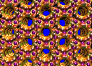
Similar to honeycombs, regular patterns of pores like those in zeolites can orderly accomodate small molecules or clusters. But if you want to entrap, say, enzymes, peptides, or large nanoparticles, you must use materials with larger pores. Some porous silicas have large honeycomb channels, while the cavities of metal organic frameworks display an amazing variety of size and shape. With those nice architectures awaiting to be filled, ordering molecules might appear like an easy task.

As you imagine, things are more complex. Perfect order cannot be achieved. All cavities would need to be uniformly occupied by the guests. This is going to be very unlikely, because molecules move a lot even when they’re confined… like bees in a hive.

front_low_res
Molecules in nanocavities are sort of like bees in a honeycomb: they form an organized colony (Artwork: Andrea Stangoni)

About bees, I had direct experience… as a child, I used to observe my dad opening up his hives to inspect them. This gave me the chance to “study” the behaviour of these awesome creatures inside their honeycomb.

Bees do not occupy all hexagonal holes in the frame, and move continuously around, without any apparent pattern. Hence they’re not perfectly ordered. In spite of this, the colony is amazingly organized, and performs an impressive number of complex tasks…. not just honey production!

Similarly, guest molecules confined in porous cages are not rigorously ordered. Yet they are organized, and the resulting host-guest materials can perform useful functions, which were absent in the free molecules. They can, for example, absorb and transfer photons like the antenna systems of plants and bacteria.

Now, the question is: can we improve the organization of the molecules and the performances of the materials? Well, first we should know how the molecules occupy the cavities, their orientation, spacing and so on. Are the guests aligned? Are they attached to the pore walls? What happens if water enters the pores? To find those answers, you should use several different techniques: each experiment will give you some pieces to compose the puzzle. And yes, computational chemistry helps a lot to figure our what happens inside the pores. Yet this remains a very difficult problem.

This is where mineral science might help.

Regular patterns of cages are very common in the mineral world. Not long ago, for example, geologists found in Antartica a mineral with the same structure of zeolite Z-SM5, a well-known and widely used artificial industrial catalyst. That was indeed a big surprise! Natural zeolites are indeed amazing: their pores contain impressively stable structures formed by small molecules and cations. Just look at this water wire:

Wchain
Water wire found in the channels of a natural zeolite

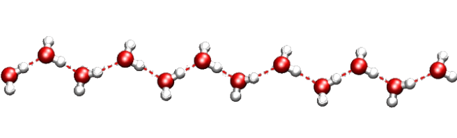
Contrary to what you’d expect, this chain is incredibly resistant to heat and pressure. First found in a rare mineral, it was named “one-dimensional ice”. But actually, our water wire “melts” at about 340 C inside the mineral framework!  This is a great example of organized structure made by Nature. You can find many others: the most famous ones are perhaps gas hydrates. Several silica minerals have hydrate structures, which are also very common in man-made porous materials. Indeed, we should pay more attention to the close links between natural and artificial host-guest materials.

Natural porous minerals, the intriguing organization of their guests, and their response to mechanical stress can be an awesome source of inspiration in the quest of more robust and efficient materials. High pressure experiments with zeolites (and also some MOF’s) have already brought us new organized materials, along with many curious facts.  But there’s so much yet to be discovered.

Perhaps, the problem with us (me included) and with our scientific era is that we don’t take enough time to relate with other disciplines. I’ve been so lucky to work with many awesome colleagues from the mineral, chemical and material science communities over the years, and it’s thanks to them that I wrote this review. One thing I learnt is that we should always try building bridges and strenghtening links between different fields because there’s nothing to lose, all to gain from a deeper exchange of ideas.

For more information….


The devil makes the pots but not the lids.

il diavolo fa le pentole ma non i coperchi

The title of this post is the literal translation of a proverb. The proverb means that Devil’s pot of wickedness sooner or later will boil – and, as there’s no lid, someone will see its content and reveal the truth. That’s the old innocent idea that, finally, justice will prevail over evil… well, I like it so much I use it as title. Rather than devils, this post is actually about pots and lids – of molecular size, of course.

As that’s not a Masterchef contest at the nanoscale, let’s get rid of the pot for the moment, and call it ‘container’. In the nanoworld there are many such containers, which can be filled with molecules. In this way, you can produce new materials with applications in various areas of technology: from solar energy to sustainability and human health.

Our containers are named zeolites – porous materials which are commonly used as adsorbents and catalysts in various commercial, industrial, and even medical applications as well as in our everyday life.  Also, if you fill zeolites with dye molecules, you’ll get materials able to capture and transfer solar energy very efficiently. You would do it much easier if you first know how their pores look like.

In particular, how do their entrances appear to an incoming molecule? This question is our “step one”,  because this information is really hard to get from experiments.

step01

Fortunately, modeling comes to the rescue…. and that’s one of the reasons why I love so much doing #compchem (computational chemistry)!!

step02

Step 2 revealed that the channel openings expose hydroxyl groups, and look somewhat like this:

zeoliteLChannel
Entrance of zeolite L channel, showing the terminal -OH groups and the channel accessibility.

Those terminal hydroxils can be condensed with other molecules, carrying specific groups, hence new properties and functionalities. Among them, the possibility of “closing” the pores. Why is it so important?

Zeolites are resistant to heat and pressure, and act as a protective shield around the dye. But every “pot” needs a “lid”:  plugging the zeolite pore entrances, so that the dyes, once included, cannot escape into the environment, would further enhance their stability.  This has already been done experimentally,  by attaching at the channel entrances peculiar molecules nicknamed “stopcocks”. They consist of two “parts”:

  • the “tail”, which can penetrate zeolite pores;
  • the “head”, which is too big to enter the pore and remains outside, thus blocking (at least partially) the channel opening.

Two typical stopcocks, one with a small tail, and the other with a long, bulkier tail, are shown below.

stopcoc

Such “molecular stoppers” do indeed a great job in preventing molecules to escape from zeolites.  However, there were no clear ideas about how these stoppers were attached to the pore entrance, and how much space they occupied.  This knowledge would help finding better “lids” for our zeolite “pots”. How do we get it? Of course by modeling, as sketched in step 3 and 4.

step03

Here’s what we learned:

  • stopper molecules prefer to bind aluminum sites at the channel entrance;
  • the tail group always penetrates inside the pore, while the head stays outside;
  • the extent of blocking depends on the stopcock.
    In particular:

     – small-tailed stopcocks are like partially opened “lids” : no full closure                – bulky-tailed stopcoks behave like “corks”: full closure

So the zeolite pore may be fully sealed by one bulky stopper, like a molecular cork on a Prosecco nano-bottle. On the contrary,  one “lid” (small stopper) leaves our “pot” partially opened. Fortunately, there’s enough room to attach a second small stopper to the opening, that can now fully be closed.

And this brings us to step 5…

step05

… which could well be the end of this story, first told some time ago. Thank you for reading it!

Anyway, there’s an epilogue, which is perhaps the nicest part (“dulcis in fundo“).  Using such information, obtained from modeling, experimental colleagues recently trapped indigo (that’s, your denim’s blue) in zeolite L, and blocked the channel entrances with two small stopcocks. In this way, they made a new pigment, exceptionally resistant, with an amazingly beautiful blue color.  For me #compchemist, that blue was simply….. the color of happiness.

zeolite_channel_openings


For more information…


How carbon monoxide binds to TiO2

What do a spacecraft, a breathalyzer, and carbon monoxide have in common? Nothing at all – you’d think. And you’d be wrong!  All three give you information on things that you cannot directly see, touch or measure. A spacecraft can capture some signal and send you beautiful images of a planet. With the help of a breath tester, a policeman may deduce the alcohol content in your blood. And using carbon monoxide, researchers may find highly reactive centers on materials surfaces.  Let’s focus on the latter and see how it works!

image1
Left image: the Soyuz spacecraft (source: Wikimedia commons). Center image: a breathalyzer (source: photograpy by Elza Fiúza/ABr, distributed under a CC-BY 3.0 license). Right image: carbon monoxide (blue=carbon; red=oxygen).

When a molecule comes in contact with surface atoms, its properties change. By measuring these changes, you get information on the surface sites interacting with the molecule.  Molecular vibrations – that you can measure by infrared spectra – provide very useful information: the vibration of carbon monoxide is very sensitive to the type of surface sites.  That’s why this molecule is used to identify active centers on catalytic materials, such as titanium dioxide.

How does carbon monoxide (CO) bind to surface atoms?  If you’re a chemistry student, you (should) know very well how CO interacts with molecules and ions. You’ve learned that this molecule can work both as a donor and as an acceptor of electron density.  Well, what’s nice, is that this happens also on surfaces, and  you can see it experimentally.

Let’s see this step-by-step. Carbon monoxide is a peculiar molecule. When it acts as a donor, charge flows to its bonding partner, which could be, for example, a metal cation.  This process strengthens the C−O bond and increases its vibration frequency.  This means that, in the infrared spectrum of the sample, you’ll find the CO band at higher frequencies – “blue-shifted” – with respect to the free, unperturbed molecule.  But carbon monoxide can also accept electron density from its bonding partner. If this occurs, the C-O bond becomes weaker: its stretching frequency decreases, and you’ll see a “red-shifted” CO band in your spectrum.

Carbon monoxide is colorless, odorless, and highly toxic – a true and unmerciful silent killer. It binds to iron(II) in hemoglobin, and this prevents the delivery of oxygen to the human tissues. This is a – very unfortunate – case where carbon monoxide acts at the same time as a donor and as acceptor. The bond is synergic: CO donates to the metal,  the metal back-donates to CO, and these two mechanisms reinforce each other:– that’s why it kills.  This synergy  occurs in many molecular complexes of transition metals and ions – often with less dangerous consequences. It’s less known on metal oxide surfaces, but it may happen as well. Is this the case of TiO2?

Not apparently, because carbon monoxide can only be a donor towards Ti cations –  they are Lewis acids, and cannot give back electron density.  The lower is their coordination number, the stronger is their acid power.  For example, a Ti cation coordinated by 4 oxygens  – Ti(4) – should be a stronger acid than one bound to 5 oxygens  -Ti(5).

Researchers use carbon monoxide to explore the activity of surface cations and deduce their environment, in particular the number of oxygen neighbors. This information connects the reactivity of a catalytic center to its molecular structure,  and may help them to improve the catalyst.  Practically speaking, they send carbon monoxide on a TiO2 sample and measure the infrared spectrum.  The rule is simple: the higher the frequency of the CO band, the more reactive are the Ti sites on the sample, and the lower their coordination number.

sitetio2
The image shows a 5-coordinated  Ti center (left) and a 4-coordinated  Ti site (right) on  anatase-TiO2 surfaces

So if you had a TiO2 sample with Ti(5) sites, and a second one with Ti(4), what would you get from the experiment?  “The second sample should show a more blue-shifted CO band, because Ti(4) is a stronger Lewis acid”.  If you answered this, you’d be wrong… because we actually did the experiment, checked with calculations, and found the contrary.  We found that CO on Ti(4) gives a less blue-shifted band – even if Ti(4) is a stronger Lewis acid.  Just as if a breathalizer estimated a lower alcohol content in a drunker driver. This could happen only if a sort of magic potion neutralized the effects of alcohol (something similar exist in real life, but it’s a mineral and belongs to the large family of zeolites). Similarly, our carbon monoxide on Ti(4) should have received an antidote against the loss of electron density. The antidote could only be electron density: but where did it come from?  Simply from the oxygen atoms bound to Ti(4): they are close enough to CO and ready to help.

In short, what happens is that CO donates electron density to Ti, but the surface oxygens donate electron density to CO. The first process strenghtens the C-O bond, but the latter has opposite effects. As a result, you find the CO signal at frequencies lower than expected.  The two mechanisms are sketched in the figure below – my attempt to explain in a simple way the two-fold nature of the Ti-CO bond on titanium dioxide surfaces.

theTiCobond2

So, if you see high frequency bands in an infrared spectra of CO, please be warned: not necessarily they are due to very reactive sites on TiO2 surfaces.  And also keep in mind that carbon monoxide gives you indirect information on your sample.  Its signal can be influenced in complex ways by several factors – you might misinterpret your data, based on simple rules. From a practical viewpoint, i think that you should be aware of this, especially if you’re working on CO, or titanium dioxide materials. More speculatively, this story might help us to better understand how molecules interact with surface atoms. The complex, delicate balance of molecular-scale interactions is at the origin of technologically important phenomena – reactivity, catalysis, photocatalysis, just to mention some of them.  Understanding these interactions more deeply could help us to improve their practical applications. Much effort is still needed, but it’s worth doing!

This research by our group has been published recently (Deiana et.al., ChemPhysChem 2016, 17, 1956; 10.1002/cphc.201600284). It was also sketched in a short summary, and by an infographics in a previous post.  Here i used other words to tell the same story, because i feel it’s important to make research results accessible to a larger community.

Titania nanoparticles, carbon monoxide and infographics.

During this weekend i tackled a challenging task: to try to explain one of my recently published papers with an infographic. My first thought was to write a blog post (maybe i’ll do it as well), but i was intrigued by the idea of lumping a couple of years of work into a few tiny lines. Although attracted by the immediacy of infographics, i never used this tool, and it sounded just the right moment to give it a go. So, i went to the Canvas site and chose a fitness club advertisement as a template. After a bit of playin’ around, that’s what i’ve got:

infografic_co_forweb4

I admit i’m quite happy with it, even if the making process was not plain sailing at all, at least for me. As a first-time user, i think that there’s room for improvement, and i’ll probably do some other attempts. Actually, i enjoyed creating this infographic!

I have published it (the infografic, i mean) in figshare (acceptance rate: 100%, publication fares: 0 €). That’s openaccess – free to download and use. Unfortunately, that’s not true for the paper – not enough funds to make it openaccess as well. Anyway, if you might want to give it a look, here’s the link:

Deiana, C., Fois, E., Martra, G., Narbey, S., Pellegrino, F. and Tabacchi, G. (2016), On the Simple Complexity of Carbon Monoxide on Oxide Surfaces: Facet-Specific Donation and Backdonation Effects Revealed on TiO2 Anatase Nanoparticles. ChemPhysChem. doi:10.1002/cphc.201600284

Another short explanation can be found here, with links to additional material.